ECVM in horses: symptoms and heritability remain in focus
ECVM – really a cause of disease? New study should bring clarity
 from Kerstin Wackermann
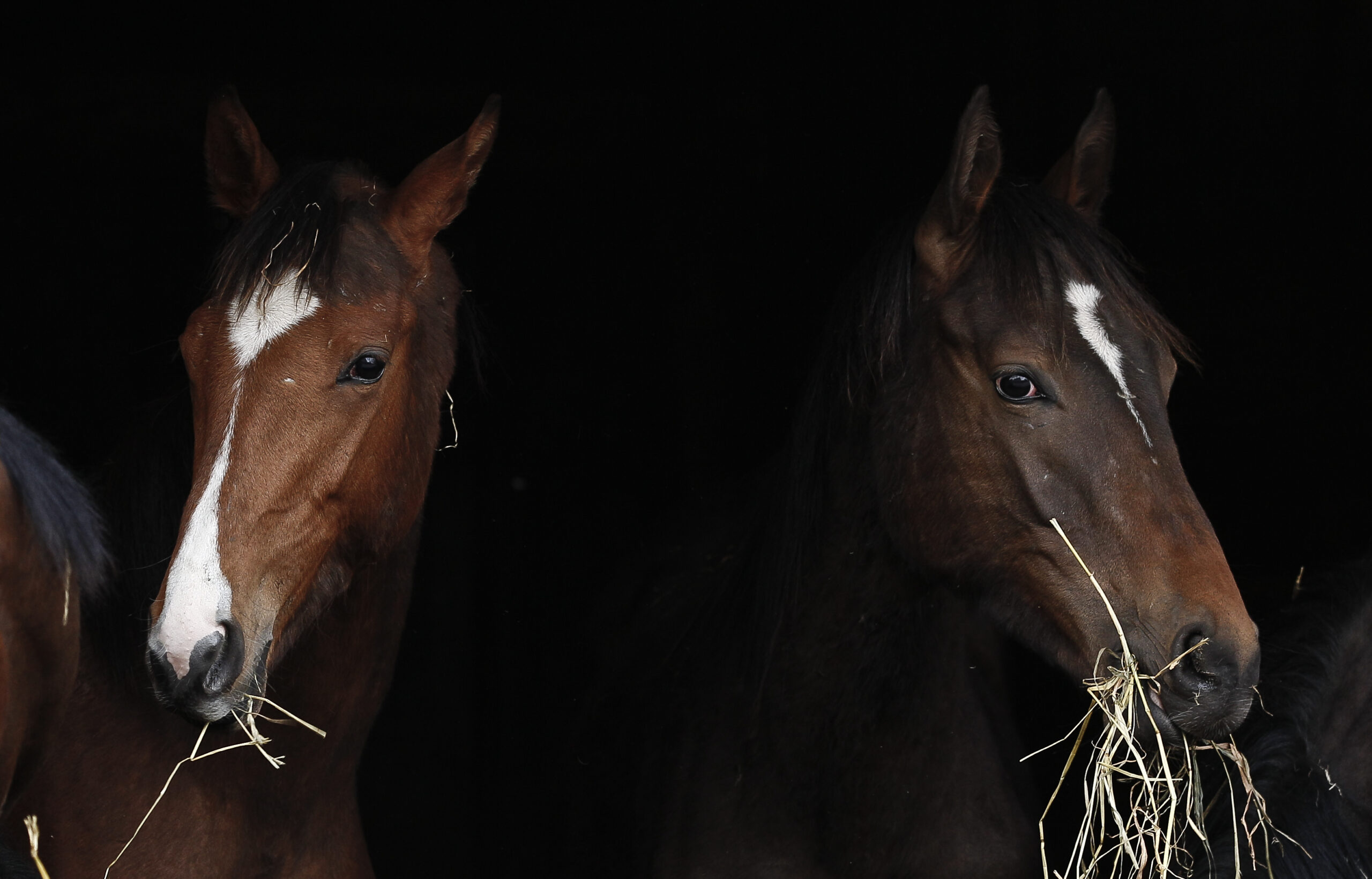
  Weakly developed neck muscles are associated with ECVM, among other things. Symbolic image: Archive Sportfotos-lafrentz.de
 The prevalence of ECVM in horses is indicated by previous study figures and the clinical experience of veterinarians. “The studies of recent years show that around 30 percent of horses have ECVM. Some vets even speak of 50 percent,” reports Dr. Arno Lindner.
The veterinarian from Jülich in North Rhine-Westphalia is a founding member of FFP e.V. and part of the EQUCAP research group. This group is currently investigating ECVM in a large-scale study – because there is still great uncertainty as to what the ECVM findings actually mean.
What is ECVM in horses?
ECVM stands for Equine Complex Vertebral Malformation. Affected horses have congenital changes to the last two cervical vertebrae. There is normally a bony plate, the lamina ventralis, on the underside of the 6th cervical vertebra. In horses with ECVM, this may be missing on the left or right side, but also on both sides, or may even be displaced to the seventh cervical vertebra. This can have consequences for the muscles that attach there. It is discussed that altered muscle attachments can lead to instability, muscle hypertrophy and pain, especially under stress or with poor training.
Changes to the first and sometimes the second rib have also been described in ECVM: The first rib can be missing in affected horses (on one or both sides) or be shorter than anatomically normal. The “than anatomically normal” is the crux of the matter: an anomaly does not automatically equate to a disease.
In its current statement, the Gesellschaft für Pferdemedizin (GPM) describes the term “Equine caudal cervical morphologic variation” (ECCMV) as more appropriate for these findings, because “malformation” already suggests a disease which, in their view, has not yet been proven for these anatomical variations.
ECVM symptoms
A central point of the current EQUCAP research project is the question of whether ECVM is associated with symptoms. Time and again, veterinarians are presented with horses that frequently stumble or suffer from coordination problems and in which the X-ray image ultimately reveals ECVM. On the other hand, there are numerous horses that remain completely normal despite the same anomaly.
“There are horses in top-class sport that show these changes on X-rays but are completely symptom-free,” explains Dr. Maren Hellige from the TiHo Hannover in the podcast “Equine Medicine Today” from the Gesellschaft für Pferdemedizin (GPM). “An X-ray finding alone is not a diagnosis. It’s just an image. And it only becomes clinically relevant when we actually have failures, i.e. when we see that a nerve is affected.” The specialist veterinarian for horses and imaging diagnostics is also part of the EQUCAP research group and is therefore working intensively on the question of whether and which symptoms are typical for ECVM.
Anyone who has been following the topic of ECVM for some time may wonder why the question of symptoms continues to be raised – after all, “classic symptoms of ECVM” have been mentioned again and again for years. Even a scientific study (Kernot et al., 2022) has compiled the following abnormalities in connection with ECVM:
– Ataxia
– pain and stiffness in the neck
– stumbling
– lameness
– wide-legged stance
– problems in body perception
– muscle atrophy in the neck
– loss of performance
– rebelliousness
– abnormal head and neck posture
The study is a systematic review, i.e. the results of previously conducted studies were summarized and evaluated. The problem from the point of view of some scientists is that although the evaluated primary studies described clinical abnormalities in horses with changes in the C5 to C7 range, they did not provide any clear evidence that ECVM is the cause of these symptoms.
Kernot et al. (2022) also point out that different imaging protocols and evaluation criteria limit the comparability of the studies. It was also unclear whether ECVM was clearly differentiated from other diseases associated with changes in the cervical spine – such as Wobbler syndrome (CVSM) – in the evaluated studies.
This is CVSM in horses
CVSM (cervical vertebral stenotic myelopathy), more commonly known as Wobbler syndrome, is a disease in which the spinal canal in the cervical spine is narrowed. This narrowing, which can be permanent (static) or occur during movement (dynamic), leads to compression of the spinal cord, resulting in neurological problems. The direct impairment of the spinal cord results in symptoms such as ataxia (coordination disorders). While CVSM is a well-researched condition, ECVM describes anatomical variations for which there is still no scientific consensus on the clinical significance.
Study from 2024: No evidence of “typical” ECVM symptoms
A more recent study comes to a completely different conclusion than Kernot et al. A research group led by Dr. Sue Dyson published the results of a cross-sectional study in 2024. Horses were examined at two clinics in England and the USA from 2017 to 2019 with the aim of finding out whether changes at C6 to T1 are associated with clinical symptoms. In this context, the researchers also looked for evidence of spinal cord compression (CVSM).
Using a defined clinical protocol, the research team examined more than 200 warmbloods. The group was divided into a “control group” (healthy horses) and a group with horses that had problems such as pain in the cervical spine, stiff gait or coordination disorders.
All horses underwent the same orthopaedic and neurological examination. This included assessing the horse’s movements in hand, on the lunge and sometimes under the rider. The neck was also palpated to detect pain reactions or movement restrictions. The researchers took X-ray images of the lower neck and the front chest area (vertebrae C5 to T2) from various angles.
It was found that the horses with problems such as stiff gait or coordination disorders were less likely to have changes at C5 to T1 than the healthy horses: 29.2% of the healthy control horses had a variant at the 7th cervical vertebra (C7). In contrast, only 16.7 % of the horses with symptoms such as ataxia or pain had this variant.
The researchers found no association between ECVM and ataxia, pain or lameness. In the horses that showed these symptoms, Dyson et al. (2024) instead frequently found other findings such as osteoarthritis of the articular processes, spondylolisthesis/ misalignment of the vertebrae, narrowing of the intervertebral foramen (through which the nerves pass) or changes to the intervertebral disc connections.
Dr. Sue Dyson, who is also involved in the current EQUCAP research project, makes a clear distinction between ECVM and CVSM. She advises looking for degenerative signs of wear and tear in horses with CVSM symptoms (ataxia). In her view, ECVM variations are often only an insignificant secondary finding.
However, the limitations of the study published in 2024 must also be noted here: the authors examined warmbloods from two referral clinics – this is not necessarily representative of the warmblood population. The study is limited to a cross-section and does not follow up the control horses. It is therefore not possible to say whether horses that were normal at the time of the study later developed clinical symptoms. The group with symptoms and the healthy control group also differed in age: the horses in the symptomatic group were on average 9.6 years old, while those in the control group were 8.5 years old.
Although oblique X-rays were taken (at an angle of 45° to 55°), these were in line with the clinical standard; a protocol specifically described for ECCMV (Gee et al., 2020) was not used, which could influence the detection of certain variants. The ethics committee had only approved images for the study that were necessary as part of a routine clinical examination anyway.
The X-ray images were each evaluated by one person at the respective clinic; a cross-clinic evaluation by several people did not take place.
X-ray on ECVM
A simple lateral image of the cervical spine is not sufficient to diagnose ECVM because the scapula and rib cage partially obscure the lower neck and the transition to the thoracic spine. Christine Gee and colleagues therefore described an X-ray procedure in 2020 that is intended to visualize variants at C6/C7 with the help of special oblique images and orientation markers. She scans the transverse processes and places a small, radiopaque marker above C5. This allows you to see on the image where C6 begins. This is intended to prevent a normal structure at C5 from being incorrectly interpreted as an ECVM variant.
In their 2023 study (which included 39 horses), Dr. Katharina B. Ros and colleagues took up the principle of oblique radiographs, but added a few points. They X-rayed C6 to T2 as well as the first and second ribs in order to detect congenital changes occurring there (known as equine cranial rib malformations, ECRM for short) using mobile X-ray equipment. The examination is carried out on a non-sedated horse, as the authors of the study believe that the horse is more stable this way: This way, the shoulder blade does not obscure T1, T2 and the first ribs so easily.
A key trick is the manual lifting of the horse’s head by 20 to 100 centimetres above the neutral position. This is particularly crucial for the depiction of ECRM, as the first ribs remain almost completely hidden in the shadow of the shoulder blade without this stretching.
For horses with massive muscles, it may be useful to carefully take back the front leg on the opposite side. The oblique images are taken at a flat angle from bottom to top (from both sides, angle range preferably 20 degrees).
This approach, complemented by a shortened imaging distance for maximum detail, reveals malformations at C7, T1 and the rib attachments that may remain hidden in a standard protocol.
In addition to the X-ray method, Dr. Katharina Ros and her colleagues developed a system for classifying the severity of the findings in their study. Such a differentiated assessment is so important because only certain degrees of malformation could actually be responsible for pain or neurological problems. The authors explain that the lack of such a grading system could be the reason why other studies have not found any clear correlations between ECVM and clinical findings.
X-ray of the cervical spine at the AKU?
Breeders, horse owners and buyers are wondering whether they should have the cervical spine X-rayed to rule out ECVM. Dr. Maren Hellige explains in the GPM podcast that X-raying the cervical spine as part of a normal purchase examination (AKU) is currently not recommended. Many findings can be made there whose clinical significance is completely unclear. The vet warns against immediately concluding that there is a disease or a problem based purely on X-ray findings.
She draws a parallel with kissing spines. For a while, it was very popular to take images of the spinous processes of the back, even during purchase examinations. In the 2018 X-ray guidelines, these X-rays were removed from the standard protocol, as it has been shown that many inconspicuous, rideable horses also show clear findings that are therefore of no clinical relevance. It has been learned that it is not the bony findings that are decisive, but the stabilization through the musculature and the correct work with the horse.
Also with regard to breeding, there is currently not enough information to advise breeders to refrain from breeding horses with certain X-ray findings on the cervical spine.
This statement corresponds to the fact that the recommended standard in the new GPM X-ray guidelines, which are to be used from April 1, 2026, does not include X-rays of the spine.
New, large-scale study on ECVM
Under the title “Investigations into the clinical significance and heritability of congenital anatomical changes of the posterior cervical and anterior thoracic vertebrae in warmblood horses”, EQUCAP has been researching since April 2024 to find out more about the symptoms and heritability of ECVM.
“We can’t compare our horse population with the horses that Dr. Sue Dyson and her team studied in California and England. It can look very different in this country. They were warmbloods there too, but the differences are still huge. We have already noticed, after around 60 horses were examined for the project, that there are differences even between the federal states,” explains Dr. Arno Lindner.
A total of between 500 and 2000 warmbloods are to take part in the research project at several German equine clinics. In order to ensure the greatest possible homogeneity of the animals studied, only closely related warmblood breeds (e.g. Hanoverian, Westphalian, Oldenburg, Holstein, KWPN) will be included in the study.
The horses are divided into different groups: 1. horses with symptoms and with changes, 2. horses with symptoms and without changes, 3. horses without symptoms and with changes and 4. horses without symptoms and without changes.
The veterinarians perform clinical and neurological examinations and take various X-ray images according to an examination protocol developed by the research group. This protocol is tailored to the specific scientific questions of the EQUCAP study and is designed to ensure that the images are of the highest possible quality for the independent reviewers (such as Sue Dyson).
The vets evaluating the X-ray images do not know whether the horse has shown clinical symptoms. This prevents “confirmation bias”: You may be more likely to see a finding in an x-ray if you know the horse is lame.
With the help of the special X-ray images and the subsequent blinded evaluation by independent experts, the research group hopes to find an objective and scientifically reliable answer to the question of the clinical relevance of ECVM.
“When we can expect the first results depends on how quickly we can examine enough test and control horses. Scientific publications are expected in 2030, perhaps 2029,” explains the veterinarian from Jülich.(Information on participation)
Is ECVM hereditary?
In addition, blood samples are being taken in the EQUCAP research project to further investigate the genetic component of ECVM. Zimmermann et al. (2023) had already provided initial indications that ECVM could be hereditary in thoroughbreds and warmbloods. The scientists had examined five skeletons of important horses.
Of these, two horses were without ECVM findings: the Hanoverian Dux and the thoroughbred Le Destrier xx. In contrast, the others showed anatomical variations: the thoroughbred stallions Der Loewe xx, Birkhahn xx and Dark Ronald xx. The malformations were very different in all three stallions, which indicates the very high variability of this trait. A total of 20 warmbloods (Hanoverian, Oldenburg, Westphalian) whose pedigrees can be traced back to these stallions were examined in the study: Of these, ten horses also exhibited malformations at C6/C7.
The family connection of the stallions studied was particularly revealing: Loewe xx and Birkhahn xx are both great-grandsons of Dark Ronald xx, which means that the study is the first to document these anatomical variations over several generations within a connected stallion line.
Another key finding of the study was that the blood percentage of Der Loewe xx was significantly higher in horses with malformations than in horses without findings. However, this result should be interpreted with caution due to the small sample size and cannot be generalized.
The researchers concluded that while this study provides initial evidence of genetic transmission, studies with much larger numbers of horses and in-depth pedigree analyses are now required to provide comprehensive evidence of the impact of influential sires on today’s population. Molecular genetic studies can provide further insights, which explains the blood sampling in EQUCAP.
Breeding associations are also researching ECVM
In addition to the EQUCAP project, another major ECVM research project has been running in Germany since 2023 under the umbrella of the International Association of Future Horse Breeding (IAFH).
The project, which is being carried out as a doctoral project at the University of Veterinary Medicine Hannover (TiHo) Foundation, involves the Nürtingen-Geislingen University of Applied Sciences and Arts (HfWU) and the Computing Center United Information Systems Animal Husbandry w.V. (vit). The IAFH also ensures a close exchange with animal breeding experts at the Universities of Göttingen and Kiel and at the Saxon State Office for the Environment, Agriculture and Geology.
The ECVM study at the IAFH also aims to clarify the clinical significance and heritability.
“Primarily warmbloods and thoroughbreds aged between two and a half and four years are being examined. The examinations will take place directly on site at rearing or training farms that are willing to participate in the project and have at least five horses of the relevant age group,” reports Gina-Lee Fischer, the doctoral student in charge, who also works as a vet and chiropractor. “So far, we have received approval for our studies in the federal states of Lower Saxony and North Rhine-Westphalia; we have applied for an extension to Mecklenburg-Western Pomerania.”
You need to know this: Applications for animal testing must be submitted for scientific studies of this kind. And as this is federally regulated in Germany, a separate permit must be obtained for each individual federal state in which horses are to be examined.
The examinations in the various rearing/training stables will be carried out uniformly by Gina-Lee Fischer. “I will be using a mobile X-ray machine and X-raying the cervical spine in accordance with the TiHo’s instructions and recommendations for X-ray diagnostics. Accordingly, the entire cervical spine will be X-rayed, including the transition to the thoracic vertebrae T1, partly T2, in order to be able to detect other possible causes of movement abnormalities. This also includes oblique images, approx. 35°, in order to be able to assess the lamina ventralis (red. note: the bony extensions of the vertebrae) on both sides.”
In order to be able to analyze the relationship between anatomical variations and clinical abnormalities more precisely, objective gait analysis using sensors is used in addition to the description when assessing movement. All horses are also subjected to a standardized orthopaedic and neurological examination.
The pedigree of the horses is only linked to the findings after all clinical and radiological examinations have been completed. This supports unbiased data collection over the entire course of the study.
The plan is to complete the data collection by fall 2026. The evaluation and subsequent publication of the results will follow immediately and can be expected from 2027.
International Association of Future Horse Breeding
The following horse breeding associations and institutions are shareholders in the IAFH: Verband der Züchter des Oldenburger Pferdes e.V. (OL), Springpferdezuchtverband Oldenburg International e.V. (OS), Trakehner Verband e.V., Holsteiner Verband e.V., Westfälisches Pferdestammbuch e.V., DSP Deutsches Sportpferd GmbH (horse breeding associations of Baden-Württemberg, Brandenburg-Anhalt, Saxony-Thuringia and Rhineland-Palatinate-Saar as well as Landesverband bayerischer Pferdezüchter e.V.), Hannoveraner Verband e.V. and Vereinigte Informationssysteme Tierhaltung w.V. (vit).
Studies on the topic
Beccati, Francesca; Pepe, Matteo; Santinelli, Ilaria; Gialletti, R.; Di Meo, Andrea; Romero, J. M. (2020): Radiographic findings and anatomical variations of the caudal cervical area in horses with neck pain and ataxia: case-control study on 116 horses. https://doi.org/10.1136/vr.105756
DeRouen, Anthony; Spriet, Mathieu; Aleman, Monica (2016): Prevalence of anatomical variation of the sixth cervical vertebra and association with vertebral canal stenosis and articular process osteoarthritis in the horse. https://doi.org/10.1111/vru.12350
Dyson, S., Phillips, K., Zheng, S. & Aleman, M. (2024): Congenital variants of the ventral laminae of the sixth and seventh cervical vertebrae are not associated with clinical signs or other radiological abnormalities of the cervicothoracic region in Warmblood horses. https://doi.org/10.1111/evj.14127
Gee, C., Small, A., Shorter, K. & Brown, W. Y. (2020): A Radiographic Technique for Assessment of Morphologic Variations of the Equine Caudal Cervical Spine. https://doi.org/10.3390/ani10040667
Kernot, N., Butler, R. & Randle, H. (2022): A Systematic Review of Clinical Signs Associated With Degenerative Conditions and Morphological Variations of the Equine Caudal Neck. https://doi.org/10.1016/j.jevs.2022.104054
Ros, Katharina B.; Doveren, Aldo; Dreessen, Christie; Pellmann, Ralf; Beccati, Francesca; Zimmermann, Elisa; Distl, Ottmar (2023): Radiological methods for the imaging of congenital malformations of C6-T1, the first and second sternal ribs and development of a classification system, demonstrated in Warmblood horses, https://www.mdpi.com/2076-2615/13/23/3732
Strootmann, T.; Peter, V.G.; Körner, J.: Radiographic Prevalence of Anatomical Variations of the Ventral Lamina of the Sixth Cervical Vertebra, C6/C7 Articular Process Joint Modeling and Competition Outcomes in Warmblood Sport Horses. Animals 2026, 16, 424. https://doi.org/10.3390/ani16030424
Zimmermann, Elisa; Ros, Katharina B.; Pfarrer, Christiane; Distl, Ottmar (2023): Historic horse family displaying malformations of the cervicothoracic junction and their connection to modern German Warmblood horses. https://www.mdpi.com/2076-2615/13/21/3415